Critical Concepts | Inside Scoop You’re Not a Guinea Pig: The Importance of Clinical Research Though negative clinical research trials like the Tuskegee Syphilis study have gained prominence over the years, a former clinical research nurse shares the value of research...
Central Line Complications and How to Fix Them
- A central line draws blood, administers fluid, and monitors hydration levels.
- Central line complications can arise, and nurses must know how to address them.
- Complications include central line-associated bloodstream infections, central venous catheter (CVC) occlusion, and catheter dislodgement.
Zunaira Rizwan
Pharm D
To inject medications, generally, we use the peripheral IV line. But in some cases, a central line is needed. Have you wondered why a central line is preferred over a peripheral line?
A central line makes it easier to draw numerous blood samples without multiple pricks, allows large amounts of fluids to be administered, and monitors the hydration status by measuring the proper atrial pressure, which helps to determine if the patient is adequately resuscitated. A central line is also beneficial when long-term intravenous therapy is required, as it can stay in place longer. After learning about the benefits of a central line, let’s dive into what it is and in what cases it is required.
What is a Central Line?
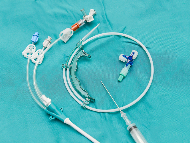
A central line, or a central venous catheter (CVC), is a long catheter used chiefly in critical care settings to access a patient’s vein. It can administer intravenous medications or total parenteral nutrition, draw blood samples, and measure central venous pressure.
A central line is inserted into a large vein, such as the internal jugular, subclavian, or femoral vein. A central line is needed for the following clinical indications:
- Vasopressor support in cases of hemodynamic instability
- Inability to acquire peripheral line access
- Needing multiple IV injections to resuscitate the patient
- Need to administer large amounts of fluids requiring a large-bore access in the vein
- Initiation of extracorporeal therapies like hemodialysis or plasmapheresis
- Need to manage central venous pressures
Central line insertion is helpful but needs proper management and utmost attention. Sometimes, various life-threatening complications can emerge because of the central line. As a nurse, you should be aware of them.
Central Line-Associated Bloodstream Infections (CLABSI)
Central line-associated bloodstream infections (CLABSI) cause most common nosocomial infections, along with hospital-acquired pneumonia. CLABSI develops within 48 hours of central line placement and is unrelated to infection at another site.
The most common organisms that cause CLABSI are coagulase-negative staphylococci, staphylococcus aureus, and Enterococcus. Methicillin-resistant Staphylococcus aureus is also seen in patients.
The signs and symptoms of CLABSI are vague, so knowing about them can be challenging. The patient may have a fever with or without chills. Assess them for signs of inflammation on the insertion site, such as redness, pain, tenderness, and swelling.
If you suspect your patient has developed CLABSI, notify the doctor. They may order a blood culture. An infected catheter may have to be removed. The healthcare provider may also request a culture of microorganisms from the catheter tip. An antibiotic sensitive to the organism should be used to treat the infection once culture results are received. According to a 2011 Centers for Disease Control and Prevention report, the mortality rate in patients developing CLABSI is 12% to 25%.
The following actions are recommendations to prevent CLABSI:
- Avoid using the femoral vein for central venous access in adult patients
- Promptly remove any catheter that is no longer needed
- Use sterile gloves to insert arterial, central, and midline catheters
- Clean the skin with >0.5% chlorhexidine preparation with alcohol before insertion of a central line
- Use sterile gauze or a sterile, transparent, semipermeable dressing to cover the catheter site
- In patients whose catheter is expected to remain in place for more than five days, use a central venous catheter impregnated with chlorhexidine/silver sulfadiazine or minocycline/rifampin
Many hospitals also provide a CLABSI care bundle to ensure policy adherence and prevent infections.
The central line can get occluded for various reasons, making it hard to flush or preventing blood from being withdrawn quickly. Occlusions can be thrombotic (due to a thrombus) or non-thrombotic (for reasons other than a thrombus.
Non-thrombotic causes of CVC occlusion include:
- Mechanical Occlusions (e.g., kinks or clamps)
- Postural Changes
- Medication Precipitate Occlusions
Thrombotic occlusions are more common than non-thrombotic ones. A thrombotic occlusion increases the risk of CLABSI, which warrants early occlusion treatment.
If you suspect an occlusion in the central line, inspect all possibilities that can cause this. Check for any kinks in the tube. See if the suture is tied too tightly to secure the central line. Ask the patient to change the posture by raising their arm or taking deep breaths to rule out postural occlusion. If the occlusion is due to a medication precipitate, consult a pharmacist. They may suggest a fibrinolytic or non-fibrinolytic agent to help clear the catheter.
Alteplase is the most common drug for thrombotic occlusions. It’s a fibrinolytic agent approved by the FDA that breaks down the clot in the catheter. Other fibrinolytic agents are also being studied.
To prevent catheter occlusions, flush all the central venous catheter lumens. Some hospitals suggest flushing the CVC with a heparin solution. The recommended amount varies but is usually between 3 ml to 5 ml. Some two-way valved devices, like the Groshong’s catheter, require only normal saline for flushing. Follow your hospital’s guidelines and flush the catheter regularly. And avoid mixing incompatible medications that may form precipitates within the CVC.
Catheter Migration or Dislodgement
Another central line complication is that the tip may migrate to another location. Flexible catheters like the PICCs are more likely to migrate. Signs and symptoms of a catheter migration include:
- Loss of blood return
- Discomfort in the upper arm during infusions
- Differing length of the external catheter from the time of insertion
If you notice that the catheter has migrated externally, do not try to push it in. However, gently pull it to the original insertion length if it has migrated internally. If the catheter is completely dislodged from its place, cover the insertion site and apply direct pressure on it. The patient will need monitoring for a possible air embolism.
To prevent a catheter from migrating, check if it is secured and the dressing is intact. Avoid pulling or manipulating the catheter. Notice the external length and compare it with the length documented at insertion.
Catheter Breakage or Rupture
Sometimes, the catheter may break due to high pressure. Small syringes like 3mL can cause increased pressure in the catheter, leading to rupture. Moreover, using sharp instruments like scissors too close to the catheter can accidentally damage or cut it.
If you notice that the catheter has ruptured or broken, clamp it near the insertion site. Place gauze over the broken area and inform the doctor. The healthcare team will then decide whether to repair or remove the catheter.
Try using larger syringes, like 10mL ones, for catheter use. These create less pressure inside the catheter than smaller ones. Avoid using sharp objects near the catheter. If you notice any occlusion and the catheter cannot be flushed, don’t force it against resistance. Doing so can result in high pressure and rupture of the catheter.
Phlebitis refers to inflammation of the veins. Mechanical and bacterial causes are central lines’ most common causes of phlebitis. Chemical phlebitis, due to a reaction to medications, can also occur but is rare. Mechanical phlebitis occurs with large catheters that are not secured in place properly.
When the catheter keeps moving, it irritates the vein. Bacterial phlebitis occurs due to bacterial infections. Failure to maintain an aseptic technique at any step can result in bacterial phlebitis.
The symptoms of phlebitis include tenderness, redness, and swelling. The patient may also have a fever if the infection is associated with a bacterial infection. Look for those symptoms to assess phlebitis.
If you think your patient has developed phlebitis, inform the doctor. The catheter may need to be removed to avoid irritation. Due to their anti-inflammatory properties, various proposed treatments, such as alternating hot and cold therapy and the application of chamomile extract, sesame oil, and marigold oil, have also been suggested.
Antimicrobial agents are administered for bacterial phlebitis but may not be enough alone. In most cases, the catheter requires removal and anticoagulant therapy. An aggressive approach like a vein resection may be needed in some cases.
A suture should adequately secure the catheter to prevent mechanical phlebitis. Nurses have also reported using cyanoacrylate glue to secure the catheter and prevent bacterial contamination.
To avoid bacterial phlebitis, follow the aseptic technique while inserting and handling the catheter. Proper handwashing, wearing sterile gloves, and using hand sanitizer all constitute essential steps in preventing bacterial phlebitis and infections like CLABSI.
Air Embolism
Air embolism is another CVC complication. The catheter can provide access to air in the vein, but it requires cautious handling by nurses. The risk of an air embolism is most significant during the insertion of a central line because of the large-bore needle. The risk increases even more when the tubing is changed, or the catheter is broken or ruptured.
The signs and symptoms of an air embolism include respiratory distress, tachypnea, and cyanosis. The jugular vein may be distended. Late signs include murmurs like a mill-wheel murmur. On auscultation, wheezing due to bronchospasm may be present. In most patients, the central venous pressure also increases.
Call the rapid response team if you suspect an air embolism. If the patient develops signs and symptoms of an air embolism while the central line is inserted, stop the procedure and clamp the line. Administer 100% oxygen to the patient and place them in the left lateral position to prevent the embolism from entering the pulmonary circulation. Consider intubation and mechanical ventilation if the patient is in severe respiratory distress.
Ask the patient to perform a Valsalva maneuver when the catheter is attached to the IV line to prevent air embolism. If the catheter breaks, clamp it near the insertion site. Flush all catheter lumens before insertion. The patient should perform a Valsalva maneuver while the catheter is being removed. After removal, cover the site with an impermeable dressing and apply pressure for five to 10 minutes.
The Bottom Line
A central line is paramount in clinical practice. In some cases, it can lead to life-threatening complications. Educate patients about infection prevention and caring for a central line. If you notice any symptoms of central line complications, inform the physician promptly. Your knowledge and vigilance toward complications that can arise and how to manage them are essential in caring for patients with a central line.
Love what you read?
Share our insider knowledge and tips!
Read More
The Nurse’s Role in Addressing Intimate Partner Violence
Critical Concepts | Inside Scoop The Nurse’s Role in Addressing Intimate Partner Violence October is Intimate Partner Violence Awareness Month, and nurses play a significant role in identifying signs of abuse. The Centers for Disease Control and Prevention reported...
What is Sundowner Syndrome?
Critical Concepts | Diagnoses What is Sundowner Syndrome? Patients with delirium or dementia can have Sundown Syndrome, a diagnosis in which emotional and behavioral changes occur beginning in the late afternoon. Mood swings, restlessness, and hallucinations can be...