Nursing Assessment
History: A detailed history can help you diagnose an aortic aneurysm. Check if the patient has a history of atherosclerosis, elevated blood cholesterol levels, hypertension, obesity, diabetes, smoking, familial tendencies, or aortic aneurysm. Ask about pain history, including its description and location.
Keep in mind the location of the aneurysm, its pain location, and type as follows:
- If the aneurysm occurs in the ascending aorta, it causes substernal chest pain that extends to the shoulder, neck, lower back, or abdomen but generally not to the jaw or arms. It is severe on the right side and described as dull, severe, and ripping pain.
- If the aneurysm occurs in the transverse arch of the aorta, it causes neck pain that radiates to the shoulders, causing a sudden and sharp tearing pain.
- If an aneurysm occurs in the descending aorta, it causes back and shoulder pain that radiates to the chest, causing sharp and tearing pain.
Check if the patient has pulmonary symptoms, such as coughing, wheezing, stridor, or dyspnea, which may be caused by descending aortic aneurysm compressing the tracheobronchial tree. Inquire if the patient has difficulty swallowing, dry cough, dyspnea, or hoarseness. All these symptoms can occur because of a transverse arch thoracic aortic aneurysm.
Physical Examination: The most common symptom of an aortic aneurysm is severe chest, neck, and back pain. Upon physical examination, the thoracic aortic aneurysm does not reveal the presence of the aneurysm itself. However, specific findings may raise your suspicion toward aortic aneurysm.
Elicit a complete neurological examination to determine the adequacy of tissue perfusion. The patient’s blood pressure should be taken from both arms because the ascending thoracic aortic aneurysm may cause changes in blood pressure readings from both arms. Also, take the pulse from the patient’s right carotid and left radial pulse and note any difference. Chest auscultation also is performed to check for pericardial friction rub and aortic valve insufficiency, which indicates the extension of an ascending aortic aneurysm proximally into the aortic valve. Monitor the patient’s pulse for bradycardia.
Assess the patient and their caregiver’s understanding of the implications of an aortic aneurysm. Assess their abilities to cope with the onset of sudden life-threatening conditions that may require prolonged hospitalization. Check the patient’s level of anxiety, knowing about their illness, potential surgery, and complications.
Since an aortic aneurysm does not cause any symptoms as such, a diagnosis is made with the help of diagnostic imaging tests that include a chest X-ray and chest CT scan. Other tests may include ECG, MRI, transthoracic echocardiography with Doppler color flow mapping, transesophageal echocardiography, and aortic angiography.
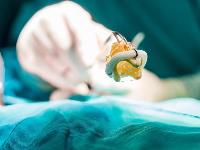
Collaborative: A thoracic aortic aneurysm measuring 4cm in size or less is often treated with oral antihypertensive agents, including beta blockers and nitroprusside. Frequent diagnostic testing is required every six months to determine the size of the aneurysm. Moreover, a thoracic aortic aneurysm having a size of 5 cm or greater is usually treated surgically. Surgical intervention is also done when the patient has intractable pain, dissection, and an unstable aneurysm that changes in size. A primary complication of thoracic aortic aneurysm is dissection. Monitor the patient for any changes in the quality of the peripheral pulses, changes in the level of consciousness, changes in vital signs, and the onset of severe, sudden, tearing, or ripping pain in the neck, back, chest, or shoulders.
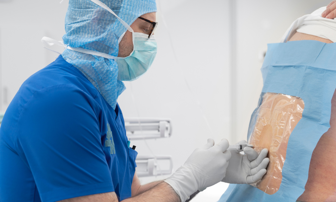
A ruptured thoracic aneurysm needs immediate surgical intervention. Before surgery, assess the patient’s peripheral pulses and monitor pulses from both sides. Take the patient’s blood pressure from both arms and auscultate for an aortic insufficiency murmur to determine the preoperative and postoperative progress.
Administer large volumes of fluids intravenously and maintain circulation until surgery is performed. The surgical procedure varies depending on the location of the aneurysm. An ascending arch aneurysm may be replaced with an interposition graft, supra coronary graft, or composite valved conduit. However, a transverse arch aneurysm is repaired with reconstructions and anastomosis. A graft helps repair the thoracoabdominal and descending thoracic aneurysms. Once the surgery is done, administer morphine or fentanyl to control postoperative pain. Also monitor the cardiopulmonary status of the patient, especially in those with congestive heart failure, because beta-blockers may worsen the preexisting congestive heart failure if the patient has elevated blood cholesterol levels that are not being controlled with a diet, a cholesterol-lowering agent is prescribed.
Independent: When an aortic aneurysm is diagnosed, it is essential to maintain adequate circulation, prevent complications, and implement patient education. If the patient does not undergo surgical interventions, educate them to consume a low-fat and low-cholesterol diet to prevent the progress of atherosclerotic plaques and prescribe statins to treat hypercholesterolemia.
Compel the patient to stop smoking and help them with smoking cessation. If they find it arduous to stop smoking, refer them to rehabilitation. For the patient undergoing surgery, focus on adequate blood circulation pre- and postoperatively. Postoperative surgical care is the same as that of patients who undergo general anesthesia, and postoperative care is similar to that of patients who undergo chest surgery. Provide aggressive pulmonary hygiene every one to two hours. Assist the patient with mobility and range of motion exercises to lower the effects of immobility. Emotional support should also be provided to the patient and their caregivers.
Discharge and Home Health Care Guidelines: Patients who do not require surgical interventions can be discharged early; however, those undergoing surgery must stay longer at the hospital. If they do not have any support at home to take care of after being discharged from the hospital, an extended care facility may be required where they can recover. Ensure that the patient and their caregivers understand all prescribed medications, including dose, route, side effects, and actions.
If the patient is overweight or obese, suggest they adhere to a low-fat and low-cholesterol diet. It is a good idea to work with a dietician who can create a customized diet plan for them that focuses on their health condition. Ensure that the patient understands why controlling blood pressure and cholesterol is imperative, as it can lead to atherosclerotic plaque progression. Encourage patients to stop smoking if they do and highlight the importance of smoking cessation. Inform them that smoking can increase the risk of hypertension and atherosclerosis.