Course
Continuous BiPAP in Respiratory Failure
Course Highlights
- In this Continuous BiPAP in Respiratory Failure course, we will learn about the types of respiratory failure and disease processes related to its onset.
- You’ll also learn mechanisms and benefits of non-invasive continuous ventilation for treating respiratory failure.
- You’ll leave this course with a broader understanding of clinical presentation of patients and assessments to evaluate baseline and response to treatment.
About
Contact Hours Awarded: 1
Course By:
Molina Allen, MSN, RN, CCRN
Begin Now
Read Course | Complete Survey | Claim Credit
➀ Read and Learn
The following course content
Introduction
Respiratory failure is due to a deficit in gas exchange within the respiratory system. There is a deviation leading to an abnormal balance between oxygen, carbon dioxide, or a combination. This may be caused by a condition that impedes air or blood flow to the lungs, alters the nerve impulses and muscle responses controlling the motion of breathing, affects the area of the brain responsible for breathing regulation, or prevents functional gas exchange within the alveoli.
Pathophysiology
Type I Respiratory Failure
Type I respiratory failure is characterized by insufficient oxygen delivery to keep up with the demands of the system’s functions. The result is hypoxemia marked by a partial pressure of oxygen (PaO2) < 60 mmHg. This may be accompanied by a normal or lowered partial pressure of carbon dioxide (PaCO2). The dysfunction may stem from the following common causes that may require non-invasive positive pressure ventilation:
Alveolar hypoventilation is an impairment of unknown cause where hypoxemia occurs in the presence of hypercarbia. This imbalance normally triggers a homeostatic response of the central nervous system to respond to changes in carbon dioxide levels.
Defects-altering diffusion occurs when there is a change in the surface area or thickness of the alveolar-capillary system that prevents normal exchange across the membrane. Since carbon dioxide more readily moves across the membrane, CO2 levels are typically normal while hypoxemia is evident. Diseases that cause anomalies leading to diffusion defects include interstitial lung disease and emphysema.
Ventilation/perfusion (V/Q) mismatch is a breakdown of the balance between alveolar ventilation and the flow of pulmonary capillary blood. A completely balanced ratio is equal or 1:1; however, a normal functional level has an approximation of 0.8 due to the difference of ventilation and perfusion shift between the lung’s apex versus bases. Higher perfusion over ventilation results in a V/Q ration of less than 1, while a right-to-left shunt is noted when the V/Q ratio attains zero. This mismatch is the underlying factor of the majority of Type 1 respiratory failure disease: COPD, ARDS, CHF, and Pulmonary Embolism.
Right-to-left shunt as described above will not improve with oxygen therapy, much like the lack of gas exchange that occurs with right-to-left intracardiac shunting. This can occur with arteriovenous malformation, severe pneumonia or pulmonary edema, and complete atelectasis.
(1)
Type II Respiratory Failure
Type II respiratory failure is defined as a rise in arterial carbon dioxide (CO2) (PaCO) of greater than 45 mmHg. The pH will be acidotic at less than 7.35. (1)
Respiratory pump failure is caused by factors that limit the mechanism that pumps blood back to the heart via inspiration. Inhalation increases the volume of the thorax due to the negative pressure pull of diaphragm contraction. With exhalation, the pressure increases.
This ultimately affects thoracic vein blood pressure, promoting the return of blood from the thoracic veins to the heart’s atria.
Increased dead space is a condition whereby the alveoli are unable to provide gas exchange. This may be due to a physical or anatomical condition. This is the most common reason that individuals with COPD are afflicted with high CO2 retention.
Increased CO production is a metabolic condition that may be related to sepsis, thyrotoxicity, exercise, or fever. This becomes problematic if respiratory compensation is not able to stabilize the pH levels. (1)

Self-Quiz
Ask Yourself...
- Why would non-invasive ventilation be more favorable to mechanical ventilation that ensures an airway is maintained?
- How would you determine which treatment would be best for the patient?
- Why is it important to determine the type of respiratory failure before initiating treatment?
- Can you give an example of when biPAP would not be advantageous to a patient?
Epidemiology
Chronic obstructive pulmonary disease (COPD) is a severely debilitating respiratory condition that overall affects geriatric populations with more severity and frequency. The projected incidence is expected to grow to become the fourth leading cause of death by 2030(2). COPD can be characterized as a systemic disease with profuse inflammation of the pulmonary system resulting in sputum production, dyspnea, and cough that progressively worsens over two weeks.
Tachypnea and tachycardia often present as symptoms increase and can lead to critical illness and need for care escalation (3). Both acute and acute chronic COPD result in hypercapnia as evidenced by PACO2 levels exceeding 45 mmHg and blood gases that are acidotic (3).
Hospitalization for respiratory failure associated with COVID-19 is required when the oxygenation demands of the system are unable to be met via the normal course of ventilation and perfusion. The type of respiratory failure that is most associated with COVID-19 is Type I; however, studies have shown that COVID-19 has complexities that compound the effects and severity of respiratory distress that are driven by a systemic response (4).
The prognosis for respiratory failure in COVID-19 to progress into pneumonia and other respiratory complications increases the probability that these patients will require supplemental oxygen at a higher rate of delivery with pulmonary support beyond that which can be offered via nasal cannula.
Early in the pandemic, mechanical ventilation was utilized as this provided respiratory support and also a closed system, which prevented viral spread, but this was found to have high instances of pneumonia and induced lung injury directly related to the use of mechanical ventilation (4).
Hypoxia that did not respond to O2 delivery at 6-16 L/min via or continuous positive airway pressure could be trialed on BIPAP as an alternative to intubation (4).
Hypocapnic obesity syndrome is due to airway resistance and a decrease in chest wall compliance. It is marked by the clinical symptom of elevated carbon dioxide blood levels in individuals with obesity while in an awake state with no other explanation for hypercapnia during the daytime (5.) Due to adipose distribution, concurrent sleep apnea, and restructuring of respiratory mechanisms related to the decreased Co2 clearance during apneic events that occur over time, acute on chronic events result in respiratory failure with hypoxic and hypercapnic status that may respond well to bi-pap treatment (5).
Pulmonary edema, also known as “flash pulmonary edema,” is a condition of the respiratory system where the pressure in the pulmonary arteries is insufficient to overcome the pressure in the alveoli. This results in fluid being siphoned from the vascular system into the lungs, causing a barrier to gas exchange (6). Heart failure is a leading cause of cardiogenic pulmonary edema, although this may also occur due to stiffening of heart valves seen with mitral valve dysfunction. Onset post-events include exposure to toxic gases, sepsis, or traumatic injury (6).

Self-Quiz
Ask Yourself...
- Some respiratory is associated with obesity, what assumptions might be made that would not be accurate with this patient population?
- The geriatric population is more prone to comorbidities and complex disease processes, how would this affect the care that you are delivering?
- You are mentoring a newer nurse who does not understand the disease process of pulmonary edema and how bi-pap improves respiratory status. How would you assist the new nurse to research and investigate while continuing to care for the patient?
Assessment
With respiratory failure, the goal is to provide supplemental support to maintain sufficient oxygenation and prevent system decompensation with developing organ failure. In both types of respiratory failure, with few exceptions, patients will require supplemental oxygen to treat acute hypoxia. Further, hypercapnia must be addressed and treated promptly to avoid CNS depression and respiratory acidosis. This triad has a high mortality rate when not identified and medically managed. To assess hypoxia and hypercapnia balance, blood gases are the most reliable laboratory result. This allows the clinician to monitor efficacy of treatment and adjust BiPAP settings for optimal respiratory therapy (7).
Pulse Oximetry is valuable to monitor treatment at the moment. This measurement of arterial oxygen saturation monitors the percentage of oxygen-bound hemoglobin. The SaO2 level should be maintained above 90% in patients without underlying, chronic respiratory issues. For patients with COPD or other respiratory diseases that alter respiratory drive, the goal is to maintain oxygen levels between 88-92% (7)
Capnometry is a non-invasive monitoring that measures the amount of carbon dioxide via exhaled air. This is a useful tool to initially assess patients in respiratory distress to evaluate CO2 retention and guide further treatment.
In identifying susceptibility of progression to respiratory failure, studies have revealed that rising levels of plasma surfactant protein D (SP-D) has a correlation with patient requiring escalation to critical care for development of acute respiratory disease (4).
LDH is another predictor of higher mortality levels, while high neutrophil-to-lymphocyte, C-reactive protein, and D-dimer were associated with less favorable outcomes (4)
Blood Gases
In Type I respiratory failure, PaO2 is low. This is a level lower than 60 mmHg. CO2 initially is not elevated. For Type II, PaO2 is low as well, with PaO2 < 50 mmHg; however, in contrast with Type I, the carbon dioxide level is also elevated. With the carbon dioxide level being elevated, the pH may be normal if compensation is adequate but would be expected to trend to acidotic of > 7.45 if left untreated or unresponsive to treatment (7).
Radiography
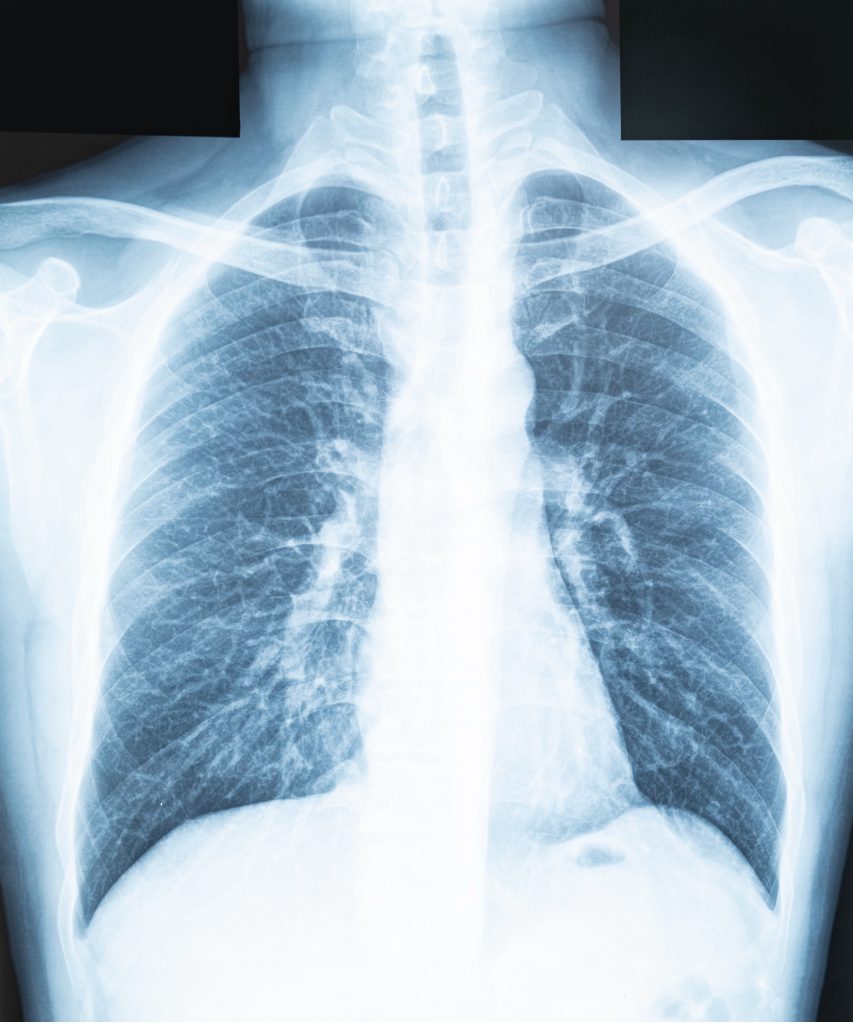
Chest imaging is a powerful tool that can be used to differentiate subtleties of severe respiratory disease. Chest X-rays are an inexpensive and quick method that is accessible in most healthcare settings to detect abnormalities of the chest wall and visualize opacities, consolidation, and fluid or air accumulation in the pleural space. The disadvantages are that in the presence of certain disease states, such as with Covid-19 and ground glass opacities, the imaging to evaluate pulmonary vessels and lung opacity will be markedly obscured (9).
CT imaging offers the most advantageous evaluation of pathological findings associated with respiratory failure; however, it does carry with it a higher dose of radiation to the patient. CT imaging, in comparison to x-ray, has increased reliability combined with higher acuity for identifying subtle nuances that may be poorly visualized otherwise. Chest x-rays do have the higher advantage of being portable to the patient in critical situations where it is difficult or would cause instability to transport and move a patient (8,9).
Clinical Signs and Symptoms
The patient presenting with acute respiratory failure will have signs of respiratory distress including tachypnea, pallor or cyanosis, diaphoresis, tachycardia, and use of accessory muscles to breathe. There may be audible wheezing, rhonchi, and grunting due to the effort being used to move the air.
Patients receiving biPAP may experience undesirable symptoms such as insomnia, lethargy, xerostomia, and the feeling of air hunger. Coughing and increased sputum production are also common during treatment (10). Anxiety is also common with both respiratory failure and with bi-pap treatment.

Treatment
Use of non-invasive positive pressure ventilation (NI-PPV) as initial treatment for respiratory failure is highly recommended by respiratory and cardiovascular organizations (4). There are two types of NI-PPV, continuous positive pressure ventilation (cPaP) and bi-pap. CPaP maintains a constant level of pressure, whereas bi-pap provides separate, titratable inspiratory and expiratory positive airway pressures. The expiratory positive airway pressure is akin to the positive end-expiratory pressure (PEEP) feature of mechanical ventilation. PEEP increases oxygenation by encouraging open airways, increasing alveolar ventilation, reinflating collapsed airways, and thus, balancing any V/Q match (13).
Bi-pap is considered the gold standard for treatment of acute respiratory failure due to pulmonary edema and acute-on-chronic COPD exacerbation. This can be delivered via nasal mask, various sizes of face mask, or full helmet. It is very important that a mask can achieve a tight seal to prevent leakage which decreases flow and pressure. Bi-pap optimizes oxygenation and tissue perfusion through improving gas exchange (4, 13).
Systemic or inhaled corticosteroids, as well as inhaled bronchodilators, may be used to decrease bronchial inflammation and prevent alveolar collapse caused by pulmonary edema. Antibiotics may be utilized to treat underlying infections and to address sepsis (12).

Self-Quiz
Ask Yourself...
- The nurse is concerned with trends of the patient’s blood gas levels; however, the provider is not responding to requests to assess the patient. How would you escalate the concern for patient safety?
- A patient is started on bi-pap due to flash pulmonary edema and a chest x-ray is ordered. Radiology requests that the patient be brought to the department for imaging. How should the nurse address this request?
- The patient’s blood gases return indicating a normalization of pH and decrease in CO2. How is this relevant to the current bi-pap treatment?
Contraindications and Complications
Contradictions
Any patient who has nausea should be closely monitored and treated promptly while bi-pap treatment is active. Precautions must be taken to prevent a full stomach, either during meal breaks if it is tolerated or if the patient is receiving parenteral nutrition. Decreased gastric emptying is common during illness, increasing the likelihood of gastric discomfort. Nausea must be treated to prevent vomiting via antiemetics to prevent aspiration from occurring due to the air pressure that would seek the path of least resistance, which would be to backflow into the lungs.
Any patient who is unconscious or has an altered drive to breathe is not suitable for bi-pap treatment due to the limited ability to protect the airway and aspiration risk. This also applies to any patient who shows signs of severe respiratory distress, such as flail chest, retractions of accessory muscles, a respiratory rate that exceeds 24 breaths per minute, or dyspnea that prevents the verbalization of a full sentence.
A tight seal is imperative to maintain for bi-pap to be effective. Facial trauma, burns, or anything that inhibits the ability to maintain a tight seal, such as intolerance of mask, is a contraindication to treatment.
Patients who are post-esophageal or gastric surgery are not ideal candidates due to the potential for tissue damage and harm to the surgical site. Any suspected or confirmed pneumothorax is also not eligible. Due to the thoracic pressure that bi-pap elicits, hypotension is common and should be addressed promptly with fluids and vasopressors, as appropriate. The systolic blood pressure should be maintained > 90.
Any altered pathway to ventilation such as a foreign body, airway obstruction, or tracheostomy eliminates the possibility to utilize bi-pap.
(11, 12, 14)

Self-Quiz
Ask Yourself...
- What is the argument for lower oxygen in disease processes such as COPD that have an altered respiratory drive?
- What effect would therapeutic communication have on a patient that is having difficulty tolerating the bi-pap mask?
- What would be some examples of patient intolerance to the bi-pap treatment?
- A patient is concerned about receiving controlled substances for anxiety. How would this best be explained to the patient of the risk for addiction?
Complications
Tension pneumothorax is a condition that may occur due to increased air pressure applied during bi-pap. For this reason, bi-pap settings should only be adjusted by respiratory or trained nursing staff. When this occurs it results in the pleural space being filled with air which prevents lung expansion due to pressure and can cause severe decreases in pressure as it limits venous return to the cardiovascular system.
This complication is identified via chest pain, tachypnea, and hypotension with mediastinal deviation on the chest x-ray. Immediate decompression is required via thoracostomy and chest tube insertion.
Anxiety is also a side effect that can be detrimental to treatment. Patients who are unable to tolerate bi-pap must be immediately intubated and mechanical ventilation initiated.
(13, 14)


Self-Quiz
Ask Yourself...
- A patient who can tolerate the mask off for short periods can intake a meal by mouth. The patient reports hunger and wants additional nutrition. How would you respond to the patient’s request?
- Discuss possible interventions to reduce anxiety that are multi-modal and include non-pharmacological options.
- What are the consequences of utilizing a bi-pap with a contraindication?
- In an emergency how would you determine whether bi-pap would be the most efficacious treatment for the patient without knowing a full history?
Nursing Care Consideration
- Patient safety with bi-pap use is extremely important, the patient must be assessed frequently for neurological status and ability to maintain their own airway. Hemodynamic monitoring is also important, and the provider should be notified immediately of any significant decrease in blood pressure or SBP under 90.
- Continuous O2 saturation monitoring with alarms as well as telemetry should be ordered to notify of concerning changes in respiratory or cardiovascular status.
- The nurse should assess for nausea and promptly treat it with antiemetics to prevent vomiting.
- Anxiolytics may be administered per order as needed to prevent combativeness and anxiety that may arise from restriction and closed-in sensation from the mask. This will assist with compliance and reduce intolerance to the mask and feeling of pressure.
- When corticosteroids are part of the treatment plan, it is important to monitor blood glucose levels for associated hyperglycemia. Sliding scale insulin may be administered as indicated to maintain euglycemia.

Self-Quiz
Ask Yourself...
- For complex patients, alarms may lead to alarm fatigue and a risk of a delayed response to an emergency. What are some ways to combat this phenomenon?
- The patient is concerned that they now have diabetes since insulin is part of the treatment plan. How could this best be explained to a patient with limited health literacy?
- How can neurological status be fully evaluated on a patient who is bi-pap dependent, and communication is limited?
- What would you do if the patient developed chest pain with accompanying hypotension?
Conclusion
Bi-pap is a non-invasive treatment that may be effective early on with several conditions that cause Type I and Type II respiratory failure. Using biPaP as the first defense, when not contraindicated, can prevent further respiratory decompensation and reduce the risks associated with mechanical intubation.
References + Disclaimer
- Mirabile, V., Shebl, E., Sankari, A., & Burns, B. (2023). Respiratory failure in adults. In StatPearls. StatPearl Publishing. Retrieved September 21, 2024, from https://www.ncbi.nlm.nih.gov/books/NBK526127/
- Radigan, K. (2023). Management of COP exacerbations in the ICU: what’s new? Critical Care [Alert], 31I (3), 17-2. Retrieved from https://www.reliasmedia.com/articles/management-of-copd-exacerbations-in-the-icu-whats-new
- Agustí A, Celli BR, Criner GJ, et al. Global Initiative for Chronic Obstructive Lung Disease 2023 Report: GOLD Executive Summary. Eur Respir J 2023; 61:2300239
- Bonnesen, B., Jensen, J., Jeschke, K., Mathioudakis, A., Corlateanu, A., Hansen, E., Weinreich, U., Hilberg, O., & Sivapalan, P. (2021). Management of COVID-19-associated acute respiratory failure with alternatives to invasive mechanical ventilation: High-flow oxygen, countinuous positive airway pressure, and noninvasive ventilation. Diagnostics, 11(12), 1-12. https://doi.org/10.3390/diagnostics11122259
- Nanayakkara, B. & McNamara, S. (2024). Pathophysiology of chronic hypercapnic respiratory failure. Sleep Medicine Clinics, 19(3), 379-389. https://doi.org/10.1016/j.jsmc.2024.04.001
- Wilson, B. (2024). Pulmonary edema. Salem Press Encyclopedia of Health. Salem Press.
- Xiong, X., & Yuan, W. (2022). Effect of noninvasive positive pressure ventilation on prognosis and blood gas level in COPD patients complicated with respiratory failure. Evidenced-Based Complementary and Alternative Medicine, 2022(3089227). https://doi.org/10.1155/2022/3089227
- Nanayakkara, B., & McNamara, S. (2024). Pathophysiology of chronic hypercapnic respiratory failure. Sleep Medicine Clinics, 19(3), 379-389. Retrieved from https://www.sleep.theclinics.com/article/S1556-407X(24)00039-0/abstract
- Meglio, L., Carriero, S., Biodetti, P., Wood, B., & Carragiello, G. (2022). Chest imaging in patients with acute respiratory failure because of coronavirus disease 2019. Current Opinion in Critical Care, 28(1), 17-24. https://doi.org/10.1097/MCC .0000000000000906
- Peterson, P., Tracy, M., Mandrekar, J., & Chlan, L. (2023). Symptoms in patients receiving noninvasive ventilation in the intensive care unit. Nursing Research, 72(6), 456-461. https://doi.org/10.1097/NNR.0000000000000688
- Pooboni, S. K. (2024). Noninvasive ventilation procedures. In Clinical Procedures. Retrieved September 25, 2024, from https://emedicine.medscape.com/article/1417959-overview?form=fpf
- National Institutes of Health. (2024). Treatment. In Respiratory Failure. Retrieved September 26, 2024, from https://www.nhlbi.nih.gov/health/respiratory-failure/treatment#:~:.text=Treatments%20for%20respiratory%20failure%20may,a%20long%2Dterm%20care%20center Lee, B., Moriyama, B., Soble, J., Diepen, S., Solomon, M., & Morrow, D. (2018). Positive pressure ventilation in the cardiac intensive unit. Journal of the American College of Cardiology, 72(13). https://doi.org/10.1016/j.jacc.2018.06.074
- Alviar, C., Miller, P., McAreavey, D., Katz, J., Lee, B., Moriyama, B., Soble, J., Diepen, S., Solomon, M., & Morrow, D. (2018). Positive pressure ventilation in the cardiac intensive unit. Journal of the American College of Cardiology, 72(13). https://doi.org/10.1016/j.jacc.2018.06.074
- Carpio, A., & Mora, J. (2023). Positive end-expiratory pressure. In StatPearls. Retrieved September 26, 2024, from https://www.ncbi.nlm.nih.gov/books/NBK441904/
Disclaimer:
Use of Course Content. The courses provided by NCC are based on industry knowledge and input from professional nurses, experts, practitioners, and other individuals and institutions. The information presented in this course is intended solely for the use of healthcare professionals taking this course, for credit, from NCC. The information is designed to assist healthcare professionals, including nurses, in addressing issues associated with healthcare. The information provided in this course is general in nature and is not designed to address any specific situation. This publication in no way absolves facilities of their responsibility for the appropriate orientation of healthcare professionals. Hospitals or other organizations using this publication as a part of their own orientation processes should review the contents of this publication to ensure accuracy and compliance before using this publication. Knowledge, procedures or insight gained from the Student in the course of taking classes provided by NCC may be used at the Student’s discretion during their course of work or otherwise in a professional capacity. The Student understands and agrees that NCC shall not be held liable for any acts, errors, advice or omissions provided by the Student based on knowledge or advice acquired by NCC. The Student is solely responsible for his/her own actions, even if information and/or education was acquired from a NCC course pertaining to that action or actions. By clicking “complete” you are agreeing to these terms of use.
➁ Complete Survey
Give us your thoughts and feedback
➂ Click Complete
To receive your certificate
