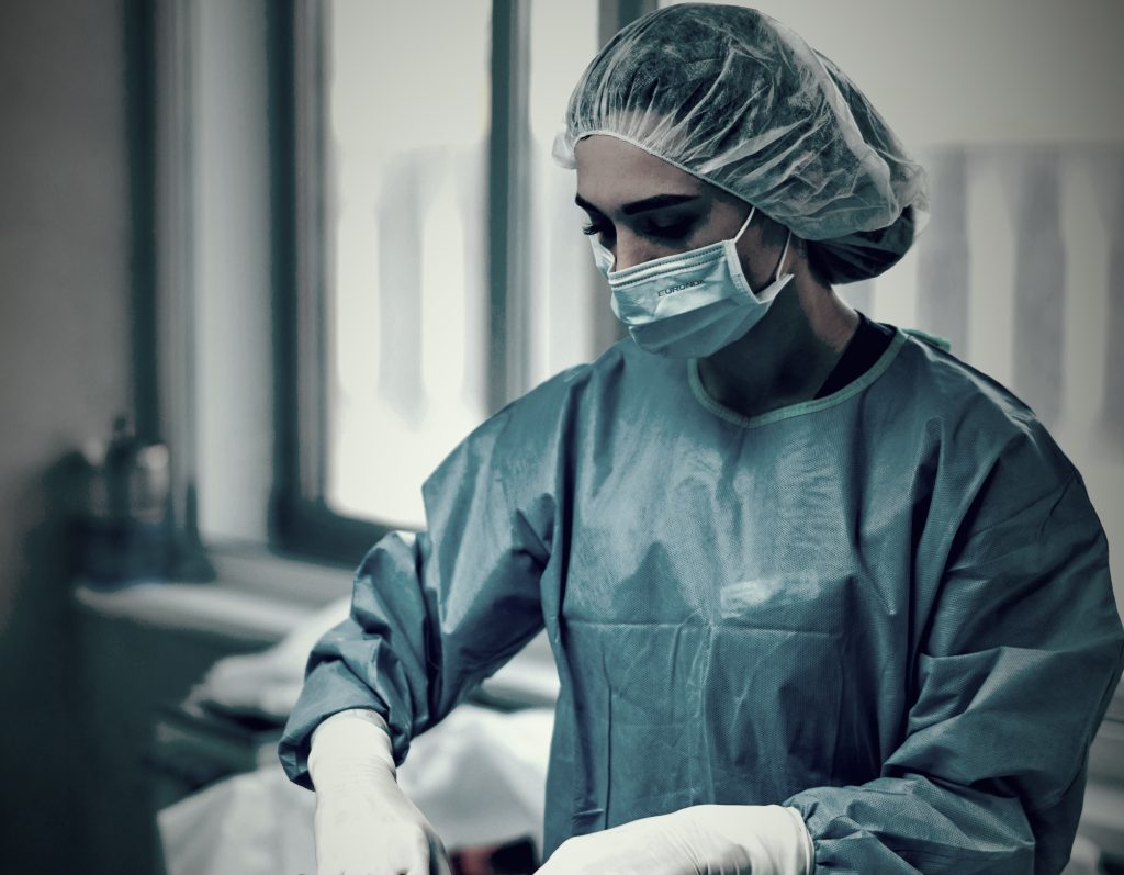
Skin Graft Care
Contact Hours: 1
Author(s):
Sarah Schulze, MSN, RN
Course Highlights
- In this Skin Graft Care course, we will learn about anatomy and physiology of the epidermis and dermis and how it relates to skin grafting
- You’ll also learn between different types of skin grafts and their indications
- You’ll leave this course with a broader understanding of contraindications for a skin grafting procedure
Introduction
Skin grafting is a surgical procedure used to treat a variety of conditions resulting in skin loss or damage. Clients may undergo skin grafting for cosmetic or reconstructive reasons, and nurses may encounter clients needing or recovering from a skin graft procedure. This is an especially relevant topic for nurses working in surgical settings or with clients suffering from burns, infection, or even skin cancer.
The nurse must have a baseline understanding of skin anatomy, how skin grafts are performed, types of conditions where grafting may be warranted, and what the healing process entails for these clients. Skin graft care is unique and requires close monitoring and regular nursing care to avoid complications and graft failure. This course will help solidify that understanding and prepare the nurse to care for skin graft clients in various settings.
Process of Skin Grafting
Anatomy and Physiology of Skin
A brief overview of the anatomy and function of the skin is necessary for understanding skin grafts. The epidermis is the thin, outermost layer of skin that primarily serves as a barrier for the body. It is largely made up of keratinocytes that produce keratin and lipids for barrier function and to enable UV absorption. The epidermis also contains melanocytes, which color skin; Langerhans cells, which have immune system function; and Merkel cells and nerve endings that facilitate touch sensation (5,10).
Beneath and connected to the epidermis is the dermis, which consists of two layers of connective tissue. The papillary layer is the thinner, upper layer consisting of loose connective tissue and blood vessels. This layer helps regulate temperature and provides nutrients to the epidermis. Dermal papillae are upward projections of dermal tissue that increase the area of contact between the dermis and epidermis for cohesive function between the two layers and play an important role in wound healing. The reticular layer is deeper, thicker, and composed of dense connective tissue and collagen fiber. This layer provides strength and elasticity to the skin and is where hair follicles, sebaceous glands, and sweat glands originate (5,10).
Ask yourself...
- How might the make-up of each layer of skin impact the grafting process?
- How would damage to various layers of skin impact healing, including the appearance and function of the skin?
Types of Skin Grafts and Indications
The location of the wound, the typical thickness of skin in that location, and the mechanical demands of that area will help determine which type of graft is best suited for the wound. The donor site should be matched to these specifications as closely as possible.
A split-thickness skin graft (STSG) involves the epidermis and a very superficial part of the dermis. STSGs are thin enough that they do not contain their own blood supply, so a healthy wound bed is necessary for the graft to survive and incorporate into the surrounding skin. This type of graft is indicated for partial to full-thickness wounds with a healthy vascular wound bed, such as with burns, trauma, infection, and ulcers. STSGs can often cover large surface area wounds, and the skin can also be meshed to increase the surface area (1, 3,5).
Skin from the abdomen, thighs, and back is often a preferred donor site for STSG. Thin grafts have low metabolic demand and typically take well and heal quickly. Thinner grafts are delicate and hold up best on body parts that do not undergo a lot of mechanical strain. Due to the thin amount of skin harvested, the healed areas may often be hypopigmented or lack hair (1,3,5)
A full-thickness skin graft (FTSG), on the other hand, consists of the epidermis and entire dermis and may be used for deeper wounds. The increased number of cells in a thicker graft has increased metabolic demand, and a healthy wound bed with adequate blood supply is necessary for the graft to adhere and heal. Healing time is longer for FTSG, and the risk of graft failure is higher.
Preferred harvest sites for FTSG often include the groin, arms, or chest. FTSG is ideal for smaller wound sites or areas of the body with high mechanical demand, like the palms of the hands. Due to the greater thickness, FTSGs may include better pigmentation and hair growth (1,3,5,6).
Less commonly, composite grafts may be used for small wounds that require some structural repair in addition to skin. Composite grafts transplant skin and other soft tissues, such as cartilage, and are commonly used to repair the nose, ears, or fingertips (1,3).
Ask yourself...
- Consider burns for an indication of skin grafting. How might the type of graft differ for a partial thickness versus a full-thickness burn?
- Which type of graft would be best used for skin loss due to frostbite? Why?
Wound Bed and Donor Site Considerations
The process of skin grafting involves transferring cutaneous tissue from one area of the body to another to help reconstruct or facilitate the healing of damaged tissue. Various levels of skin thickness can be used, each with its own implications for appearance, blood supply, and healing.
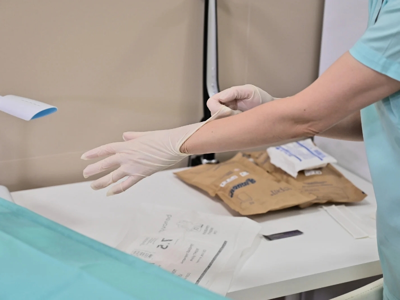
When preparing a client for skin grafting, several areas require consideration. First is the wound itself and the location and condition of the wound bed. For a graft to successfully adhere to a wound bed, the wound must consist of a base of healthy tissue with good vascularization. Non-viable tissue or any exudate should be debrided to prepare the wound site. A clean wound bed and edges are paramount to a successful graft (1,3,4).
The donor site must also be considered. Appropriate donor sites must be chosen considering both the suitability of the harvested skin for the graft and the ability of the donor site to heal well, as it will be a new wound. The size and thickness of the area to be harvested, surgical positioning, ease of accessing both sites and aesthetic match (such as skin color and texture) of the wound and donor sites should all be weighed when determining where to harvest from.
Flat regions with large surface areas, such as anterior lateral thighs, the abdomen, the back, and lateral arms and legs, are commonly chosen donor sites. Areas that may be easily covered by clothing are often selected for aesthetic reasons. For clients with extensive burns, donor sites may be limited and need to be chosen from healthy, unaffected areas (1,3,4).
Procedure Details
During a grafting procedure, the wound bed must be cleaned and debrided. The donor site must be selected and cleaned as well. For larger or irregularly shaped grafts, a template of the wound is often made to ensure the right size for transfer. For FTSG, the donor site is excised using a scalpel, and any adipose tissue may be trimmed off the graft. For STSG, the skin is lubricated with mineral oil, and an electric surgical tool called a dermatome is used to shave off a thin layer of skin (1).
The graft is placed in a sterile saline solution while the donor site is tended to for hemostasis and wound dressing. Then, the client is repositioned for graft placement, ideally with very little time between excision and placement (1).
If a graft needs to be expanded to fit a larger area, meshing may be used. This can be done manually or with a meshing device that creates perforations in the skin that allow it to be stretched and expanded to cover a larger surface area. The more meshing applied to a skin graft, the longer it will take to heal, as the graft must fill in the perforations between skin bridges. There is some benefit to the holes in a meshed graft, as they can serve as drainage holes for exudate and blood to drain rather than collecting between the wound bed and graft (1,4).
Whether the graft is meshed or not, once it has been excised and the wound bed is properly prepared, it can be applied by firmly placing it over the wound bed. The edges are sutured in place, and a bolster or pressure dressing is applied to keep the graft from shifting. Appropriate wound care over the next two weeks will help heal both the newly grafted wound site and the donor site (1,2).
Ask yourself...
- Why is the thickness and condition of the wound site important before a client undergoes skin grafting?
- Why might it be difficult to harvest adequate grafting tissue from a burn victim?
- What are the benefits of meshing a skin graft? What are the risks and drawbacks?
Outcomes
Under ideal circumstances, a healthy wound bed will nourish the skin graft while new, fragile vasculature forms between the wound and the graft. As the graft begins to be supplied with blood flow, integration should occur, and epithelial tissue will fuse the 2 parts together (also filling in any holes from meshing). The appearance, sensation, and function of the skin graft should provide better results than allowing the area to heal by secondary intention, which often results in large amounts of scar tissue and lack of innervation and temperature regulation (1,3,4).
The new blood supply for the graft should be established by 5-7 days of healing, though it will take several weeks of pressure dressings and wound care before the area fully integrates. For donor sites, FTSGs will take 2-3 weeks to heal due to the thicker graft excision, and STSGs will take around 1-2 weeks. After initial integration, it can take up to a year before the maturation process of the graft is complete, including more uniform pigmentation and softening of the skin. Some scarring at both the graft and donor site is expected (1,3,4).
Risks and Complications
As with any treatment option, skin grafting has risks. Graft failure is the greatest risk and can occur with varying prevalence (anywhere from 3% to 30%) depending on the specific details of the individual case. After the initial transfer, the wound bed nourishes skin grafts while revascularization occurs.
If the graft is separated from the wound bed or its metabolic demands are too great for the wound bed to support, revascularization will be interrupted or not occur, and the graft will fail, allowing the tissue to die. Graft separation can occur from shearing force from improper wound treatment or client activity level or from fluid or hematoma formation between the graft and wound bed. Meshing may reduce the risk of this complication (1,5).
Signs that a graft may be failing to revascularize include pallor, gray discoloration, or even black eschar around 1-2 weeks after grafting. Some superficial sloughing or necrosis may take place even a few weeks after the grafting, even if healthy tissue is developing underneath, so proper wound care is necessary to distinguish between the two (1,5).
Additional risks include contracture or deformed healing, especially for STSG over irregularly shaped wounds, keloids or excessive scar formation, infection, chronic pain, loss of sensation, skin discoloration or lack of hair growth, and scarring all of which can occur at the graft site as well as the donor site (1,5).
Of course, there are conditions where skin grafting is contraindicated altogether for client safety or high risk of graft failure. A skin graft should absolutely not be performed for wounds that are actively infected or where bleeding has been unable to be stopped. Clients who have had skin removed due to cancer but the removal was incomplete should also not undergo a skin graft (1,5).
Clients who smoke, have a bleeding disorder, take anticoagulant or long-term corticosteroid medications, or are malnourished are at an increased risk for graft failure and poor wound healing and are poor candidates for the procedure (1,5).
FTSGs should only be used in small areas (<1cm) if the wound bed is avascular. An STSG for wounds without clear margins is at an increased risk of puckering and contracture (1,5).
Ask yourself...
- Which phase of the healing process is the most important for a successful graft? Why?
- Consider the client population you most often work with. If needed, who would be a good candidate for skin grafting? Who would be at high risk of graft failure?
- What are the risks of skin grafting unique to a client with a full-thickness burn down to the bone?
- Why is a client taking blood thinners at an increased risk of graft failure?
Nursing Care
Nursing care of clients at any stage of the skin grafting process requires an understanding of the procedure and the graft’s healing and integration process. Nurses must provide direct care to clients during the acute stages of graft integration, including wound care, dressing changes, and pain control measures. Nursing education during acute healing and longer-term care is also paramount to optimum outcomes.
Protecting the Graft
First and foremost, nurses must protect the graft site to ensure proper integration and prevent graft failure. Extreme care when changing dressings, ambulating clients, and providing client care must be used in order to prevent shearing the fragile new vasculature that is forming in the first few days and weeks after graft transfer. Counseling clients on careful position changes and movements, depending on the location of the graft, is important. Clients should wear the prescribed dressing at all times and avoid soaking in water. The nurse should thoroughly review a client’s medication list and ensure the provider knows any medications that increase bleeding risk, as these may need to be discontinued. At the same time, the graft first heals (9).
Dressing Changes and Wound Care
Skin grafts may have different types of dressings depending on the location and thickness of the graft. A bolster is a dressing that is stitched to the surrounding tissue to hold the graft in place. This is not typically removed until the graft has integrated over the next 1-2 weeks. Pressure dressings may also be used to apply pressure to the site and keep fluid and blood from building up between the graft and wound bed, as well as keep the graft from shifting. Pressure dressings may be held in place with sutures or a splint. Gauze or dressing pads may also be used, typically over the top of a bolster or on the donor site (1,2,9).
Dressings should be checked for drainage, exudate, and blood frequently while clients are in the hospital, and removable dressings should be changed if they are saturated at any point. Dressings should be routinely removed and changed, and the wound should be assessed every 2-3 days during the initial weeks. The wound should be kept clean and dry as much as possible, and dressing changes are a great time to assess the wound for signs of revascularization, infection, or graft failure (1,2,9).
Lotion or ointment to keep the skin moisturized and healing well may be applied as directed by a provider, typically after the initial integration.
Assessing the Graft
Both the graft and the donor site should be assessed frequently. The graft should be examined for signs of infection, bleeding, ischemia, and graft failure. Assessments such as purulent exudate, obvious shifting of the graft, bleeding, hematomas, eschar, and necrosis should be reported to the provider immediately. Some mild edema is common in the first few days, but excessive swelling or redness should be addressed. The graft may appear pink at first, but over time, the color should resemble that of the surrounding tissue. Graft color should not be pale or gray, which indicates inadequate blood supply and possible graft failure.
The donor site needs to be assessed for signs of infection and bleeding as well (1,2,9).
Pain Control
Clients may experience pain from both the wound and the donor sites. Different facilities and providers may have different protocols and options for pain management in the first few days and weeks after a graft, ranging from continuous subcutaneous anesthetic injection to the application of ice, topical anesthetics, and hydrocolloid wound dressings. Clients may also be given oral opioid and non-opioid medications for pain control, with the intensity and duration of pain being highly dependent on the nature of the initial injury/wound and the size of the graft (1,8,9).
Clients should be assessed for pain and the efficacy of pain management frequently, as often as every 1-2 hours in the first few days. Pain should improve steadily as healing occurs, and intense or unrelenting pain may be a sign of infection and need further evaluation (1,8,9).
Education
Clients should be advised not to engage in strenuous activity for several weeks, avoiding bending or lifting, depending on where the graft is. Grafts on dependent limbs should be elevated to reduce swelling and pain.
As dressings are removed (usually after the first 3-4 weeks) and grafts are open to the air, clients should avoid clothing that rubs or irritates the area and avoid scrubbing in the shower. Daily skin care in the form of moisturizers and SPF for UV protection is necessary for long-term wound healing and reduction of scarring. Clients should avoid smoking, drinking alcohol, or using recreational drugs, as these can all slow wound healing. In general, donor sites will heal quickly and more easily than graft sites. Clients should also be assessed for self-esteem issues stemming from the appearance of grafts and donor site scarring (7,9).
Ask yourself...
- Why is a bolster or pressure dressing more important for skin grafts than other types of wounds?
- Why would a dressing saturated with blood indicate a need for immediate assessment of a grafted wound?
- What do you think is the top nursing priority for a client with a skin graft?
- Why is long-term skin care important for best results with a skin graft?
Case Study
Let’s consider the case study of a 45-year-old male client who presents to the ED with a partial thickness burn to 50% of the left lateral thigh following a cooking accident at home involving hot oil. The decision is made to perform a split-thickness skin graft (STSG) for optimal wound healing and to minimize scarring. The client has no significant medical history, is a non-smoker, and has no history of diabetes or vascular disease.
The opposite thigh was selected for the donor site due to its similar skin color and texture. Given the partial thickness of the burn, a split-thickness graft is taken using a dermatome and then run through a meshing device. The graft is placed carefully onto the debrided burn wound, ensuring full coverage, and is secured with a bolster dressing.
Postoperatively, the client is closely monitored for signs of graft adherence and potential complications. Dressing changes are scheduled every 2–3 days, during which the wound is inspected for signs of infection, hematoma, and graft failure. Special care is taken to prevent infection or mechanical disruption of the graft.
Dressing changes are uneventful, and the wound and donor sites appear healthy. Small areas of serous drainage are noted, requiring additional attention during dressing changes, but signs of infection do not develop. Pain management is achieved with non-opioid analgesics, and the client reports minimal discomfort at both the graft and donor sites.
By the end of the second week, the graft has fully adhered, and the bolster dressing is removed. Mild hyperpigmentation is present, but the client is reassured that skin coloration will improve over time. The client is educated on the importance of long-term care, including applying moisturizers and sun protection to the graft site to prevent dryness, discoloration, and excessive scarring. The donor site heals rapidly within 10 days, showing minimal scarring.
Overall, the client’s recovery has been uneventful. The STSG has proved effective in closing the wound and promoting functional skin regeneration.
Ask yourself...
- What risk factors does this client have for undergoing a skin graft?
- What client factors make him a favorable candidate for a skin graft?
- Based on this client’s injury, why do you think a STSG was chosen?
- What purpose does the bolster dressing serve in this client’s case?
- What assessment data indicates this client’s wound is healing well?
- What assessment would you expect for poor wound healing or signs of graft failure?
- What do you know about long-term skin graft maturation that makes moisturizer and SPF important for client education?
Conclusion
Skin grafting is a useful treatment option for clients experiencing skin damage or loss from infection, burns, ulcers, or other causes. For a graft to be successful, significant care must be taken to promote revascularization, optimum healing, and graft maturation. By being well-informed about skin anatomy, the grafting process, and acute and long-term graft care, nurses can play an important role in promoting proper wound healing. Completing this course should provide the necessary foundation for quality nursing care.
References + Disclaimer
- Braza ME, Fahrenkopf MP. Split-Thickness Skin Grafts. [Updated 2023 Jul 25]. In: StatPearls [Internet]. Treasure Island (FL): StatPearls Publishing; 2024 Jan-. Available from: https://www.ncbi.nlm.nih.gov/books/NBK551561/
- Cartier, A., Barbier, M. A., Larouche, D., Morissette, A., Bussières, A., Montalin, L., Beaudoin Cloutier, C., & Germain, L. (2022). Tie-Over Bolster Pressure Dressing Improves Outcomes of Skin Substitutes Xenografts on Athymic Mice. International journal of molecular sciences, 23(10), 5507. https://doi.org/10.3390/ijms23105507
- Cleveland Clinic (2021). Skin graft. Cleveland Clinic. https://my.clevelandclinic.org/health/treatments/21647-skin-graft
- Elseth A, Nunez Lopez O. Wound Grafts. [Updated 2022 Oct 31]. In: StatPearls [Internet]. Treasure Island (FL): StatPearls Publishing; 2024 Jan-. Available from: https://www.ncbi.nlm.nih.gov/books/NBK564382/
- Prohaska J, Cook C. Skin Grafting. [Updated 2023 Aug 16]. In: StatPearls [Internet]. Treasure Island (FL): StatPearls Publishing; 2024 Jan-. Available from: https://www.ncbi.nlm.nih.gov/books/NBK532874/
- Ramsey ML, Walker B, Patel BC. Full-Thickness Skin Grafts. [Updated 2023 Jul 24]. In: StatPearls [Internet]. Treasure Island (FL): StatPearls Publishing; 2024 Jan-. Available from: https://www.ncbi.nlm.nih.gov/books/NBK532875/
- Schulz, A., Rothermund, I., Lefering, R., Fuchs, P. C., & Schiefer, J. (2018). Long-term Scar Quality after Treatment of Standardized Partial-Thickness Skin Graft Donor Sites. Advances in skin & wound care, 31(3), 109–117. https://doi.org/10.1097/01.ASW.0000527287.28216.65
- Sinha, S., Schreiner, A. J., Biernaskie, J., Nickerson, D., & Gabriel, V. A. (2017). Treating pain on skin graft donor sites: Review and clinical recommendations. The journal of trauma and acute care surgery, 83(5), 954–964. https://doi.org/10.1097/TA.0000000000001615
- Workman, D.I.M. L. ([Insert Year of Publication]). Medical-Surgical Nursing (10th ed.). Elsevier – Evolve. https://pageburstls.elsevier.com/books/9780323654050. Pgs 437-472
- Yousef H, Alhajj M, Fakoya AO, et al. Anatomy, Skin (Integument), Epidermis. [Updated 2024 Jun 8]. In: StatPearls [Internet]. Treasure Island (FL): StatPearls Publishing; 2024 Jan-. Available from: https://www.ncbi.nlm.nih.gov/books/NBK470464/
Disclaimer:
Use of Course Content. The courses provided by NCC are based on industry knowledge and input from professional nurses, experts, practitioners, and other individuals and institutions. The information presented in this course is intended solely for the use of healthcare professionals taking this course, for credit, from NCC. The information is designed to assist healthcare professionals, including nurses, in addressing issues associated with healthcare. The information provided in this course is general in nature and is not designed to address any specific situation. This publication in no way absolves facilities of their responsibility for the appropriate orientation of healthcare professionals. Hospitals or other organizations using this publication as a part of their own orientation processes should review the contents of this publication to ensure accuracy and compliance before using this publication. Knowledge, procedures or insight gained from the Student in the course of taking classes provided by NCC may be used at the Student’s discretion during their course of work or otherwise in a professional capacity. The Student understands and agrees that NCC shall not be held liable for any acts, errors, advice or omissions provided by the Student based on knowledge or advice acquired by NCC. The Student is solely responsible for his/her own actions, even if information and/or education was acquired from a NCC course pertaining to that action or actions. By clicking “complete” you are agreeing to these terms of use.
Complete Survey
Give us your thoughts and feedback!