Course
Time is Brain: Door-to-Needle Time
Course Highlights
- In this course, learners will review different types of strokes and learn how to identify them
- The importance of early recognition, testing, and treatment to decrease disability and mortality will be discussed
- Learners will leave this course with a better understanding of “Door to Needle Time” and be able to apply this understanding in clinical practice
About
Contact Hours Awarded: 2
Course By:
Nellie Chinyanta
BSN-RN, CEN, MPH
Begin Now
Read Course | Complete Survey | Claim Credit
➀ Read and Learn
The following course content
In this course, we will discuss the importance of early recognition of stroke symptoms in improving the quality of care and client outcomes. When it comes to strokes, time is the brain. The more time goes on without treatment, the higher the risk of disability and potential mortality. As a healthcare provider, you must recognize what interventions are needed immediately after diagnosis.
Introduction
The clinical definition of a stroke is “syndrome of acute, focal neurological deficit attributed to vascular injury…of the central nervous system”(15, 22). A stroke, also known as a brain attack, is a medical emergency that occurs when there is a disruption of blood flow to the brain (15, 22). This can be caused by an occlusion of one of the blood vessels within the brain or an intracranial hemorrhage (bleeding within the brain) (15, 22). When either occurs, brain tissue is deprived of oxygen and nutrients, which, if prolonged, can lead to tissue death (4, 22).
A stroke due to an occluded vessel is called an ischemic stroke, and one caused by bleeding within the brain is known as a hemorrhagic stroke (4). Approximately 85% of all strokes are ischemic strokes. They are caused by atherosclerosis (plaque buildup within the vessels) or an embolus (a blood clot that travels through the blood and causes an obstruction) occluding blood flow within one of the vessels in the brain (16). There are rare instances where strokes are caused by vascular malformations, brain aneurysms, and cerebral cavernous malformations (24).
There is a third type of stroke, known as a transient ischemic attack (TIA), which occurs when blood clots occlude blood flow within a vessel but break up before any lasting damage happens, with complete symptom resolution within 24 hours (24). However, people who experience TIA are at higher risk of having an actual ischemic stroke than those who have never had a TIA (24). Therefore, a client presenting symptoms of a TIA should have all the recommended stroke testing and treatments to help mitigate their stroke risk (24).
In the US, over the past few decades, mortality rates due to stroke have steadily decreased, and this is mainly attributed to advancement in early recognition and treatment of acute strokes (16). However, strokes are still the leading cause of death worldwide and the leading cause of acquired long-term disability (9). The high incidence of strokes is caused by a mix of socioeconomic factors and a high prevalence of modifiable risk factors (8).


Self-Quiz
Ask Yourself...
- What is the clinical definition of a stroke?
- What are the three most common types of strokes?
- What is the direct impact of disrupted blood circulation on brain tissue?
- How would you explain to a client the difference between a TIA and an acute ischemic stroke?
Types of Strokes
The most common type of stroke in the US is ischemic stroke, which occurs when a blood clot or plaque embolus travels to the brain and cuts off circulation to a particular area (24). Some of the hallmark symptoms of ischemic strokes are as follows (4,15).
- Sudden onset of unilateral (one-sided) weakness in the face or extremities.
- Sudden onset of altered mentation, difficulty speaking or understanding speech (aphasia and dysarthria).
- Sudden disturbance in vision
- Sudden difficulty ambulating, loss of balance, lack of coordination (ataxia or vertigo).
- Sudden onset of a severe headache.
Hemorrhagic strokes are less common, accounting for approximately only 13% of all strokes in the US (24). This type of stroke occurs when a blood vessel in the brain ruptures and bleeds into surrounding brain tissue. The human skull does not expand; thus, when a bleed happens, the affected brain tissue becomes compressed (24). There are two subtypes of hemorrhagic strokes: intracerebral hemorrhages occur within the brain, and subarachnoid hemorrhages occur between the inner and outer layers of the tissue covering the brain (23).
Three main conditions increase the lifetime risk of hemorrhagic stroke, these are (9,20, 24).
- Hypertension—Uncontrolled high blood pressure can weaken blood vessels over time and cause arteries within the brain to rupture.
- Cerebral aneurysm: A weakened spot develops on the arterial wall of a brain vessel. As blood flows past this spot, the pressure leads to an outpouching of the arterial wall (an aneurysm). As the outpouching grows, the walls become weaker and eventually rupture.
- An arteriovenous malformation is a congenital condition that involves an abnormal tangling of arteries and veins in the brain that bypasses capillaries. Sometimes, these can rupture and lead to a hemorrhagic stroke.
The presenting symptoms of a hemorrhagic stroke are sometimes similar to those of an ischemic stroke and can also include.
- A sudden, intense headache (often referred to as the “worst headache of my life”)
- Double or blurry vision
- Nausea and/or vomiting
- Loss of consciousness (or decreased level of consciousness)
- Sudden onset of seizures
- Neck stiffness
Over the years, the acronym Balance, eyes, face, speech, time (BE FAST) has emerged as a community education guide and prehospital clinical tool for rapidly recognizing acute stroke symptoms (4,15,17). Acute onset of aphasia, dysarthria, ataxia (dizziness or vertigo), changes in level of consciousness or alertness, or a sudden headache onset can indicate that someone is experiencing an acute neurological emergency (5,17).
Self-Quiz
Ask Yourself...
- List 3 hallmark stroke symptoms
- What would be your first suspicion in a client who is present with the worst headache of their life?
- Can you list the symptoms of stroke represented in the BE FAST stroke mantra?
- What are the three conditions often associated with an increased risk for hemorrhagic stroke?
Epidemiology
According to the CDC, strokes are the 5th leading cause of death in the United States and also the leading cause of long-term disability (5). However, it is noteworthy that the incidence of stroke in the US for both males and females has significantly decreased since the 1990s (5). While strokes are possible in people from all backgrounds and of all ages, they are more prevalent among specific demographics and in the presence of particular risk factors. The primary modifiable risk factor for strokes is hypertension, defined as a blood pressure reading equal to or greater than 130/80 mmHg (5). The following is a non-exhaustive list of non-modifiable stroke risk factors.
- Age: Stroke risk also increases with age, with the highest incidence occurring among people 65 and older.
- Gender Also plays a role, as men generally have a higher overall risk than women.
- Women of childbearing age have an added risk during pregnancy or with the use of hormonal birth control pills.
- Pre-existing health conditions: Presence of health conditions that increase stroke risk, including hypertension, diabetes, heart disease, and obesity.
- Race-African Americans have the highest overall risk of developing a stroke within their lifetime. While Hispanic Americans and Indian/Alaskan natives have a higher risk than white Americans, they have a lower lifetime risk for developing a stroke than their African American counterparts.
- 1 in 365 African Americans has the genetic disorder Sickel cell disease, which can also lead to strokes.
- 3 in 5 American Americans have a diagnosis of obesity, diabetes, or high blood pressure, which all increase one’s lifetime risk of a stroke.
- Other health conditions include family or personal history of a previous stroke or TIA, genetic clotting or vascular disorders, and sickle cell disease.
In addition to non-modifiable risk factors, there are modifiable lifestyle factors that increase one’s risk of a stroke. The probability of a stroke increases with the number of risk factors present. Hypertension remains the primary modifiable risk factor that can lead to strokes (5,19,24). Modifiable stroke risk factors include.
- Physical inactivity, being overweight or obese, high cholesterol
- Excessive alcohol intake, cigarette smoking, and drug abuse
- Hormonal birth control pills.
A transcontinental study by Hankey researched which factors are associated with stroke and their statistical significance in increasing the likelihood of a stroke (5). They found ten modifiable risk factors, which are
- Hypertension-defined as a blood pressure more significant≥140/90
- A sedentary lifestyle or lack of regular physical activity
- High apolipoprotein levels (proteins that bind to lipids to form lipoproteins, which transport lipids in the blood, lymph, and cerebrospinal fluid)
- Diet-unhealthy cardiovascular diet
- A higher waist-to-hip ratio
- Psychosocial factors-depression, poverty, lack of access to adequate healthcare
- Cigarette smoking
- Diagnosis of cardiac disease
- High levels of alcoholic consumption
- Personal history of diabetes.

Self-Quiz
Ask Yourself...
- What is the primary modifiable risk factor for strokes?
- What lifestyle changes would you teach your client being discharged after a TIA?
- Can you list three non-modifiable risk factors?
- Your client is 37 y/o F who was diagnosed with a TIA; what risk factors particular to women of childbearing age would you educate her on?
Time is Brain
“Time is brain” was first coined in the 1990s by Dr. Camilo Gomez to describe the time-sensitive nature of stroke intervention procedures (9). Neurologists estimate that, on average, 1.9 million neurons, 14 billion synapses, and 7.5 miles of myelinated fibers are destroyed each minute during a large vessel occlusion (LVO) event (9). With this in mind, an astute practitioner does well to be adequately prepared to care for a client suspected of having an LVO and do everything they can to expedite treatment for clients who qualify for intravenous clot-busting medication or endovascular surgery (1,4,21).
Case Study
You are a nurse working in a Level 2 comprehensive stroke center and receive an EMS call at 5 p.m. letting you know they are bringing in a 60-year-old male who was found on the floor by his wife 10 minutes ago. According to the client’s wife, the client was last seen well at 9 a.m. this morning, but she spoke to him on the phone 30 minutes ago, and he sounded normal. Their wife reports that when she found him on the floor, his speech sounded funny, and he could not get off the ground even with assistance, which was not normal for him.
On EMS arrival, the client had slurred speech and a left-sided facial droop and was unable to move his left arm and leg. His initial blood pressure was 220/110, his heart rate was 80, and he is breathing 12 times a minute; his current GCS is 15. The client has a history of high blood pressure and is not on blood thinners. They are 10 minutes out. They could not obtain IV access, so they were given one dose of sublingual nitroglycerin to check blood pressure. On arrival at the ER, the client was alert and oriented x4; GCS was now 14, and the client was now lethargic. EMS stated that this change happened en route.
The stroke team is activated pre-arrival, and on arrival at the ER, the client is immediately taken to CT for imaging of his head to rule out an acute stroke. The physician orders a completed CT scan, and the client is transferred to a room for further evaluation. His BP is still elevated with a current reading of 196/105, HR is 80, responding only to painful stimuli, GCS is now 6-he is not opening his eyes, pupils are fixed and dilated, and there are no verbal responses, and he is withdrawing from pain.
He is intubated to help protect his airway and is started on a Nicardipine infusion to control his blood pressure. The radiologist reads CT imaging results within 30 minutes, and they show an extensive intracerebral hemorrhage. Given the location of the bleeding and the risks associated with endovascular surgery, the client’s family chose to enact comfort measures for the client rather than pursue aggressive treatment.
As explained above, stroke identification and treatment are very time-sensitive as brain tissue death happens quickly, and the probability of significant disability or death increases with time. Two primary stroke interventions are time-sensitive; thus, establishing a client’s last known well (LKW) is an essential part of their assessment and preparation for treatment (4, 7,8). For clients eligible to receive TPA, the treatment window is up to 4.5 hours from symptom onset; for those with an LVO, the treatment window is extended up to 24 hours from symptom onset. Even with those set definitions, clients who receive treatment closer to their LKW have better overall outcomes (6,8,9).
Self-Quiz
Ask Yourself...
- Based on the above scenario, what medical emergency may this client be experiencing?
- What are the most common presenting symptoms of an acute stroke?
- What is the most common modifiable stroke risk factor that is evident in the above case study?
- What are some of the neurological assessments that should be performed to catch acute changes in neurological status?
Emergency Stroke Management Terminology
To effectively understand the conversation around acute stroke interventions, it is essential to list out and define the important terms related to “Time is Brain” within the acute care setting. The following are the key terms around stroke identification and treatment: the last known well (LKW), door to needle time (DTN), large vessel occlusion (LVO), TPA, and thrombectomy (aka endovascular surgery).
The last known well (LKW) refers to the last time a client was known to be in their regular state of health with no acute neurological symptoms (13). Determining a client’s LKW is very important because it can determine what neurological interventions they may qualify for (8,9,11). This also undermines the importance of the “time is brain” philosophy because the more time since the onset of symptoms, the higher the chance of increased brain tissue damage (6,13).
The second important term that needs to be defined is large vessel occlusion (LVO). An LVO is the obstruction of large proximal cerebral arteries, often resulting in significant neurological deficits (10, 11). LVOs account for approximately 15-30% of acute ischemic strokes (AIS) in the United States, and they carry a higher risk of morbidity and mortality than other types of ischemic strokes (11, 14,24). Non-modifiable factors that predispose people to higher risks of ischemic strokes are race, ethnicity, and age (1,10,24).
One of the most effective treatments for an AIS in eligible clients is a minimally invasive procedure called endovascular thrombectomy (6,12). Around 2015, endovascular thrombectomy emerged as a preferable treatment option for eligible clients over medical management (8,11,24). During an AIS, blood flow to a targeted area of the brain is severely limited, causing brain tissue to undergo time-dependent irreversible damage.
Scientists and medical professionals have developed a theory known as the reperfusion hypothesis that during an AIS, there is an area of brain tissue known as the core (the area directly affected by the stroke) and the penumbra (an area of at-risk tissue proximal to the core. The reperfusion hypothesis states that the penumbra can be rescued from damage by swift restoration of blood flow, which can be achieved by timely mechanical thrombectomy (6,8). Similarly, if a client is experiencing a hemorrhagic stroke, timely interventions to stop the bleeding and restore blood flow can reduce the neurological damage from a stroke (10,24).
The term thrombolytic therapy (thrombolysis) refers to using a medication to destroy blood clots and/or prevent new ones from forming (16). To date, tissue plasminogen activator(tPA) is the standard for AIS treatment and is also the only FDA-approved medication for thrombolysis as a treatment for AIS (16). This medication can only be given within 3-4.5 hours of symptom onset, and thus, clients who may present with a stroke and have an unknown last known well (LKW) time or LKW greater than 4.5 hours do not qualify for this treatment (8).
Self-Quiz
Ask Yourself...
- What is the significance of a client’s LKW in determining their treatment options?
- Your client has been diagnosed with an ischemic stroke; you explain to his family that his type of stroke is caused by what mechanism of action?
- What is the significance of time in managing acute stroke clients?
- How would you explain the method of action of tPA in treating AIS to your client?
Rapid Stroke Screening
A stroke assessment is incomplete without a discussion of clinical screening assessments. In the prehospital setting, the Cincinnati Prehospital Stoke Scale (CPSS) is the standard; it assesses the following factors (26): presenting symptoms, rapid diagnostic imaging, and GCS.
- Facial drift
- Arm drift
- Slurred speech
The National Institutes of Health Stroke Scale (NIHSS) and vision, aphasia, and neglect (VAN) are the standard assessments in the hospital setting. The NIHSS is a more detailed assessment that looks at 15 neurological categories, and one can score anywhere from 0 to 42, with a score of 21-42 indicating a severe stroke. The NIHSS assesses the following categories (21).
- Level of consciousness
- Vision
- Extraocular movements
- Facial palsy
- Limb strength
- Ataxia
- Sensation
- Speech and language
The VAN assessment is focused on cortical symptoms of LVO, and while it has a similar sensitivity to the NIHSS, it is often used in conjunction with NIHSS because it has better symptom specificity and overall accuracy (21).
Self-Quiz
Ask Yourself...
- What are the two acute care stroke assessments used in the hospital setting?
- List 4 neurological categories that are examined in the NIHSS.
- What is the advantage of using the NIHSS and VAN assessments for AIS clients?
Door To Needle Time
In 2010, the American Heart Association launched a quality improvement campaign to improve acute stroke care for clients eligible for treatment with the clot-busting medication tPA (15). The initiative’s name was Target Stroke; this initiative included encouraging hospitals to achieve a goal of at least 50% or more of all their eligible AIS clients receiving TPA within 60 minutes of arrival at the hospital (14). To achieve this target, the AHA also established guidelines that can be condensed down to the following.
- EMS pre-notification of hospitals for incoming AIS clients
- Activating the stroke arrival team with a single call
- Rapid triage protocol and stroke team notification.
- Rapid acquisition of brain imaging-Door to CT scan time of less than 25 minutes.
- Rapid interpretation of brain imaging-CT scan read and reported by a radiologist within 45 minutes of arrival to the ED.
- Rapid laboratory testing of glucose levels and clotting times (INR, PT/PTT) for clients on blood thinners. Results should be available within 30 minutes of arrival to the ER.
- Use of specific protocols and tools to reduce delays
- Premixing tPA so the medication is ready as the decision is made to treat
- Rapid performance data feedback (14).
For AIS clients, the following evidence-based metrics measure a hospital’s success in adhering to and implementing the AHA guidelines.
- IV thrombolysis: arrive by 3.5 hours, treat by 4.5 hours: Percentage of people who come to the hospital within 3.5 hours of their LKW and receive treatment with tPA within 4.5 hours of their LKW.
- Early antithrombotics: Percentage of clients with an AIS or TIA who receive antithrombotics by the end of hospital day 2.
- VTE prophylaxis: Percentage of clients with an AIS, hemorrhagic stroke, or stroke not otherwise specified who receive VTE prophylaxis the day of or the day after hospital admission.
- Antithrombotics: Percent of clients with an AIS or TIA who receive a prescription for antithrombotic therapy at discharge.
- Anticoagulation for AFib/Aflutter: Percent of clients with an AIS or TIA with atrial fibrillation/flutter discharged on anticoagulation therapy.
- Smoking cessation: Percent of clients with an ischemic or hemorrhagic stroke, or TIA with a history of smoking cigarettes, who are, or whose caregivers are, given smoking cessation advice or counseling during hospital stay.
- Intensive statin: Percent of AIS or TIA clients discharged on intensive statin therapy.
The measure for evaluating hospitals’ quality and efficiency in managing acute stroke clients is known as door-to-needle time (DTN or DTNT) (14). The DTN refers to the amount of time between a client’s arrival at the hospital (Emergency Room) and the time clot-busting medication tPA is injected into their veins (15). Research has demonstrated a direct correlation between DTN in-hospital mortality, long-term mortality, symptomatic intracranial hemorrhage (ICH), and hospital readmission rates (6).
Researchers found that clients with longer DTN times (within 90 minutes of hospital arrival) had higher rates of all-cause mortality and higher rates of hospital readmission within 1 year (6). Therefore, the push to achieve shorter DTN times cannot be understated based on the long-term health impacts of delays in treatment (16). Reducing DTN time requires a multifaceted approach and collaboration between first responders, emergency room teams, and the clinical stroke team. Zhang et al. found that one of the keys to reducing DTN is implementing a system for sharing real-time information that starts with pre-notifying team members of a potential AIS client’s arrival and efficient communication across the clinical team (20).
In 2015, the AHA expanded its campaign to set guidelines for the use of endovascular therapy (EVT) in eligible clients (15). According to the 2015 guidelines, the AHA recommends a door-to-device time of 90 minutes of arrival to the ER and a goal of 60 minutes from door to device for clients being transferred in from a different facility (15). As stated earlier, faster intervention times can lead to better client outcomes and reduced severe morbidity and mortality rates (15).
Case Study
A 61-year-old female presents to ER triage with her family at 08:00 a.m. They stated this morning she could not speak or hold anything with her R hand. Pt has a history of high blood pressure and is not on thinners. Her LKW was last night at 11 p.m. Pt is triaged, and based on her symptoms, she is taken to CT, where a head and neck CT is performed. Once back in her ER room, she is placed on the monitor, and her EKG shows she is in Afib with an HR of 150; her BP is elevated at 195/100, other vital signs are stable, and GCS is 12-pt is unable to speak, but is making incomprehensible sounds.
Within 20 minutes, the CT results are read and show the client has an LVO; since the client’s LKW was less than 24 hours ago, the patient is a candidate for mechanical thrombectomy. The client is started on medications to control her heart rate and blood pressure and is prepared for IR. She has a successful thrombectomy and is transferred to ICU; on arrival to ICU, she is AA, OX, GCS is 15, and she is moving all her extremities.

Self-Quiz
Ask Yourself...
- Based on the presenting symptoms, what type of stroke is the client likely experiencing?
- How long is the LKW window for a client to qualify for IR thrombectomy?
- What is the target door to CT time under the Target Stoke initiative?
- Within what timeframe must a CT for hemorrhagic stroke rule be read and interpreted by a radiologist?
Diagnostic Testing
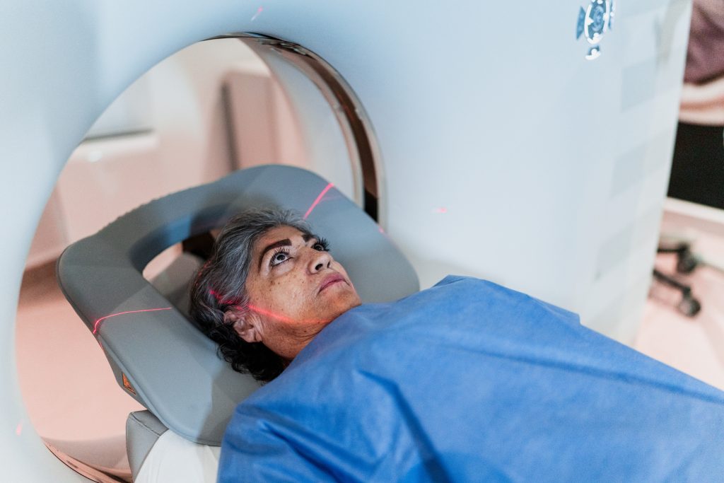
Computed tomography (CT) is the standard initial diagnostic imaging method and the most widely used method to identify a stroke (2,9,13). Some of the factors that make non-contrast CT the first imaging of choice include its rapid ability to detect brain bleeding, relatively quick study, and wide availability as a method of evaluation (2,12).
During the initial imaging evaluation, a non-contrast CT is followed by a CT angiography (CTA) of the head and neck, which is used to identify intracranial large vessel occlusions, cervical carotid, and/or vertebral artery diseases (2). Due to their high sensitivity, wide availability, and relatively fast interpretation, CTs and CTAs are the fastest methods to identify clients who are candidates for stroke intervention (2).
Other imaging options that are sometimes used as part of the evaluation for a stroke workup include magnetic resonance imaging (MRI) and magnetic resonance angiography (MRA) (2). However, these are more useful for projecting long-term prognosis and attempting to understand the source of a stroke (2).
Case Study
You received a call about a 51-year-old male client coming in via EMS with c/o R-sided facial droop, R arm weakness, sudden onset of a terrible headache, and intractable vomiting. The LKW was 30 minutes ago when he was at the table with his wife. The hospital stroke team is activated before the client’s arrival, and the client is taken straight to CT upon arrival at the ER.
After his imaging, the client is taken to the room. During his triage, you note that his current GCS is 7 (he is not opening his eyes, has no verbal response, and localizes to pain). After 15 minutes, the client’s head CT results have been read, and how the client has a hemorrhagic brain bleed that is operable. The client’s wife is at the bedside and gives consent for EVT; the client is intubated and transferred to IR for definitive treatment. Two days post-op, the client is A, OX4, GCS 15; pt is forgetful and has some residual R-sided weakness but no other focal neurological deficits.
Self-Quiz
Ask Yourself...
- Based on his symptoms, what type of stroke do you suspect this client is having?
- What is the goal door-to-device time for clients requiring EVT?
- List 5 AHA recommendations for improving outcomes for stroke clients
- The client’s wife mentioned that she feels everything is being rushed and everybody is in a hurry; what rationale would you give her for why timing is important?
Future Considerations
As mentioned above, current data has demonstrated improved stroke management trends over the past decades. However, there remains room for improvement in how acute care centers manage stroke clients. One retrospective study found that the median DTN is 65 minutes, averaging 49-88 minutes (10). This delay directly correlates with higher mortality and readmission rates (10). More studies have, therefore, called for an emphasis on reducing the benchmark DTN from 60 to 30 minutes (16). One study found that clients who received tPA within 30 minutes or less of arrival to an acute care facility had shorter hospital stays and better NIHSS scores than clients with DTN times of 45-60 minutes (16).
While tPA is currently the only FDA-approved medication for IVT, a newer medication known as Tenecteplase (TNK) is emerging as a more efficient treatment for AIS (5). TNK is a fibrinolytic alternative to tPA and has an off-label use as a treatment for acute stroke (5). There is evidence of shorter DTN times and no greater risk of an intracranial hemorrhage than those for tPA (5). In addition, compared to tPA, which is given as a one-minute bolus dose followed by a 60-minute intravenous infusion, TNK is provided as a single dose treatment over 5 seconds (5). AIS treatment aims to minimize brain tissue damage by reducing the time from the onset of symptoms to reperfusion (5).
Another stroke initiative is increasing the percentage of LVO clients receiving intravenous thrombolytic therapy within 30 minutes of hospital arrival and endovascular thrombectomy (8, 16,19). A study by Man et al. showed that with every 15-minute increase in DTN, there is a higher probability of never being discharged home, less time at home among those discharged home, and higher mortality rates (16,19). Overall, timely IVT clients had better long-term health outcomes and lower mortality rates (19). This underscores the importance of focused process improvement initiatives to minimize delays in DTN.
Improving DTN by default means improving eligible clients’ CT and EVT referral times. Therefore, hospitals must strive to create clinical pathways that expedite AIS diagnosis and treatment times (25). Reducing the incidence of strokes and reducing the morbidity and mortality associated with strokes requires a multifaceted approach that includes public health stroke education initiatives, improved first responder clinical pathways, and effective communication. Advances in medicine and technology will also contribute to improved DTN times.
Self-Quiz
Ask Yourself...
- What is the advantage of reduced DTN?
- When you prepare a presentation to your team on improving efficiency in your stroke protocol, you teach them that longer DTN times are associated with what negative long-term health outcomes?
- One of your newly graduated team members asks you why it matters if the tPA is started 15 minutes late; what evidence-based explanation would you give them?
- What is the primary advantage of TNK over tPA in reducing DTN?
Conclusion
Time is brain is more than just a slogan; it is the guiding principle for acute stroke management. The more extended brain tissue is deprived of oxygen and other essential nutrients, the higher the probability of irreversible tissue damage. The tissue damage manifests itself in more significant neurological deficits and higher mortality and morbidity rates. Faster door-to-needle times and door-to-device times are credited with lower hospital readmission rates, less long-term disability due to the effects of the stroke, and lower overall mortality rates. Effective community education on rapid recognition of the signs and symptoms of an acute stroke and the importance of timely treatment remain the keys to reducing the incidence of strokes worldwide.
References + Disclaimer
- De Havenon, A., Zaidat, O. O., Amin-Hanjani, S., Nguyen, T. N., Bangad, A., Abbasi, M., … & Al Kasab, S. (2023). Large vessel occlusion stroke due to intracranial atherosclerotic disease: identification, medical and interventional treatment, and outcomes. Stroke, 54(6), 1695-1705.
- Czap, A. L., & Sheth, S. A. (2021). Overview of imaging modalities in stroke. Neurology, 97(20_Supplement_2), S42-S51.
- CDC. (2024, April 25). Stroke facts. Stroke; CDC. https://www.cdc.gov/stroke/data-research/facts-stats/index.html
- El Ammar, F., Ardelt, A., Del Brutto, V. J., Loggini, A., Bulwa, Z., Martinez, R. C., … & Goldenberg, F. D. (2020). BE-FAST: a sensitive screening tool to identify in-hospital acute ischemic stroke. Journal of stroke and cerebrovascular diseases, 29(7), 104821.
- Hall, J., Thon, J. M., Heslin, M., Thau, L., Yeager, T., Siegal, T., … & Siegler, J. E. (2021). Tenecteplase improves door‐to‐needle time in real‐world acute stroke treatment. Stroke: vascular and interventional neurology, 1(1), e000102.
- Hendrix, P., Sofoluke, N., Adams, M. D., Kunaprayoon, S., Zand, R., Kolinovsky, A. N., Person, T. N., Gupta, M., Goren, O., Schirmer, C. M., Rost, N. S., Faber, J. E., & Griessenauer, C. J. (2019). Risk Factors for Acute Ischemic Stroke Caused by Anterior Large Vessel Occlusion. Stroke, 50(5), 1074–1080. https://doi.org/10.1161/STROKEAHA.118.023917
- Hollist, M., Morgan, L., Cabatbat, R., Au, K., Kirmani, M. F., & Kirmani, B. F. (2021). Acute stroke management: overview and recent updates. Aging and disease, 12(4), 1000.
- Jadhav, A. P., Desai, S. M., & Jovin, T. G. (2021). Indications for mechanical thrombectomy for acute ischemic stroke: current guidelines and beyond. Neurology, 97(20_Supplement_2), S126-S136.
- Karceski, S. (2021). Treating stroke from inside the blood vessel: time matters. Neurology, 97(22), e2250-e2252.
- Man S, Xian Y, Holmes DN, et al. Association Between Thrombolytic Door-to-Needle Time and 1-Year Mortality and Readmission in Clients With Acute Ischemic Stroke. JAMA. 2020;323(21):2170–2184. doi:10.1001/jama.2020.5697
- Montaño, A., Hanley, D. F., & Hemphill III, J. C. (2021). Hemorrhagic stroke. Handbook of clinical neurology, 176, 229-248.
- Negrotto, M., & Al Kasab, S. (2021). Evidence on Mechanical Thrombectomy in Acute Ischemic Stroke. 12 Strokes: A Case-based Guide to Acute Ischemic Stroke Management, 3-18.
- Nicholls, J. K., Ince, J., Minhas, J. S., & Chung, E. M. L. (2022). Emerging Detection Techniques for Large Vessel Occlusion Stroke: A Scoping Review. Frontiers in neurology, 12, 780324. https://doi.org/10.3389/fneur.2021.780324
- Parikh, N. S., Merkler, A. E., & Iadecola, C. (2020). Inflammation, autoimmunity, infection, and stroke: epidemiology and lessons from therapeutic intervention. Stroke, 51(3), 711-718.
- Phase III Higher Goals For Greater Good. (2018). In www.heart.org. American Heart Association: Target Stroke . https://www.heart.org/en/-/media/Files/Professional/Quality-Improvement/Target-Stroke/Target-Stroke-Phase-III/TS-Phase-III-5-6-19/FINAL5619-Target-Stroke-Phase-3-Brochure.pdf?sc_lang=en
- Rajan, S. S., Decker-Palmer, M., Wise, J., Dao, T., Salem, C., & Savitz, S. I. (2021). Beneficial effects of the 30-minute door-to-needle time standard for alteplase administration. Annals of clinical and translational neurology, 8(8), 1592–1600. https://doi.org/10.1002/acn3.51400
- Rioux, B., Brissette, V., Marin, F. F., Lindsay, P., Keezer, M. R., & Poppe, A. Y. (2022). The Impact of Stroke Public Awareness Campaigns Differs Between Sociodemographic Groups. Canadian Journal of Neurological Sciences / Journal Canadien Des Sciences Neurologiques, 49(2), 231–238. doi:10.1017/cjn.2021.76
- She, R., Ge, J. & Mei, Z. New Wine in an Old Bottle: tPA for Ischemic Stroke Management. Transl. Stroke Res. (2023). https://doi.org/10.1007/s12975-023-01209-6
- Target: Stroke – When Seconds Count. (2024). Www.heart.org; American Heart Association. https://www.heart.org/en/professional/quality-improvement/target-stroke/learn-more-about-target-stroke
- Teleb, M. S., Ver Hage, A., Carter, J., Jayaraman, M. V., & McTaggart, R. A. (2017). Stroke vision, aphasia, neglect (VAN) assessment—a novel emergent large vessel occlusion screening tool: a pilot study and comparison with current clinical severity indices. Journal of neurointerventional surgery, 9(2), 122-126
- Vinding, N. E., Butt, J. H., Lauridsen, M. D., Kristensen, S. L., Johnsen, S. P., Krøll, J., … & Fosbøl, E. L. (2023). Long-term incidence of ischemic stroke after transient ischemic attack: a nationwide study from 2014 to 2020. Circulation, 148(13), 1000-1010.
- Walter, K. (2022). What is an acute ischemic stroke? Jama, 327(9), 885-885.
- Xian, Y., Xu, H., Smith, E. E., Saver, J. L., Reeves, M. J., Bhatt, D. L., … & Fonarow, G. C. (2022). Achieving more rapid door-to-needle times and improved outcomes in acute ischemic stroke in a nationwide quality improvement intervention. Stroke, 53(4), 1328-1338.
- Yale Medicine. (n.d.). Hemorrhagic Stroke. Yale Medicine. https://www.yalemedicine.org/conditions/hemorrhagic-stroke
- Zhang, Y., Zhu, Y., Jiang, T., Liu, J., Tang, X., & Yi, W. (2023). An in-hospital stroke system to optimize emergency management of acute ischemic stroke by reducing door-to-needle time. The American Journal of Emergency Medicine, 69, 147-153.
- Zohrevandi, B., Monsef Kasmaie, V., Asadi, P., Tajik, H., & Azizzade Roodpishi, N. (2015). Diagnostic Accuracy of Cincinnati Prehospital Stroke Scale. Emergency (Tehran, Iran), 3(3), 95–98.
Disclaimer:
Use of Course Content. The courses provided by NCC are based on industry knowledge and input from professional nurses, experts, practitioners, and other individuals and institutions. The information presented in this course is intended solely for the use of healthcare professionals taking this course, for credit, from NCC. The information is designed to assist healthcare professionals, including nurses, in addressing issues associated with healthcare. The information provided in this course is general in nature and is not designed to address any specific situation. This publication in no way absolves facilities of their responsibility for the appropriate orientation of healthcare professionals. Hospitals or other organizations using this publication as a part of their own orientation processes should review the contents of this publication to ensure accuracy and compliance before using this publication. Knowledge, procedures or insight gained from the Student in the course of taking classes provided by NCC may be used at the Student’s discretion during their course of work or otherwise in a professional capacity. The Student understands and agrees that NCC shall not be held liable for any acts, errors, advice or omissions provided by the Student based on knowledge or advice acquired by NCC. The Student is solely responsible for his/her own actions, even if information and/or education was acquired from a NCC course pertaining to that action or actions. By clicking “complete” you are agreeing to these terms of use.
➁ Complete Survey
Give us your thoughts and feedback
➂ Click Complete
To receive your certificate